Oral cancer is easier to treat when it is found early. Research shows that about half of cases are found at an advanced stage, when treatment is harder and survival drops.
Your dental visit is one of the few times a trained professional looks closely at the soft tissue inside your mouth.
AI for oral cancer detection is helping dentists analyze photos and other clinical data to flag areas that may deserve a closer look.
The dentist still reviews every finding, and only a biopsy can confirm cancer. Think of AI as a second set of eyes that supports the screening process rather than replacing the dentist.
Most of these tools are still being studied, and results in research do not always carry over to everyday offices. Knowing what AI can and cannot tell you helps you ask better questions and act quickly if a spot needs follow-up.
Key Takeaways
- Dentists look for white, red, or non-healing spots that could be early warning signs.
- AI can flag suspicious areas in photos, but a dentist decides what happens next.
- A biopsy and histopathologic examination are used to confirm a suspected oral cancer diagnosis.
What Early Changes Are Dentists Looking For?
Dentists look for color changes, rough patches, and sores that do not heal, especially in people with known risk factors.
These early changes are often painless, which is why a routine exam catches what you might miss at home.
Which Oral Lesions Need a Closer Look?
During an oral cancer screening, your dentist checks your lips, cheeks, gums, tongue, the floor and roof of your mouth, and your neck.
They are looking for oral mucosal lesions, a clinical term for any unusual spot on the lining of your mouth.
Some spots belong to a group called oral potentially malignant disorders. These are changes that carry a higher chance of turning into cancer over time. The common ones include:
- Oral leukoplakia: a white patch that cannot be wiped off and has no other clear cause.
- Erythroplakia: a red, velvety patch. It is less common than leukoplakia but more concerning.
- Oral submucous fibrosis: a stiffening of the mouth lining, often tied to chewing betel quid, that can make it hard to open wide.
Other warning signs include a sore that lasts longer than two weeks, a lump, bleeding without a clear reason, or numbness.
One scoping review in PubMed Central lists white or red lesions, non-healing sores, and trouble chewing or swallowing among common symptoms, and names leukoplakia as an early sign.
In practice, most spots turn out to be harmless, such as a cheek bite or irritation from a denture. Your dentist will often recheck a spot in about two weeks to see whether it heals.
Who May Benefit from More Frequent Screening?
Your risk goes up with certain habits and exposures. Tobacco use in any form (cigarettes, cigars, pipes, chew, or snuff) is the leading cause.
Heavy alcohol consumption raises risk on its own, and the risk climbs sharply when you also smoke.
Human papillomavirus (HPV) is linked mainly to cancers at the back of the throat, including the base of the tongue and tonsils.
Age and sex play a part too, with risk rising as you get older and men affected more often than women.
If any of these apply to you, tell your dentist. They may suggest checking your soft tissues at every dental cleaning and exam and taking photos of any spot so changes can be tracked over time.
How Does AI Assist During an Oral Cancer Screening?
Technology can support screening, but a careful clinical examination remains essential.
At All In The Family Dental, oral cancer screening gives the dental team an opportunity to examine the soft tissues of your mouth and identify unusual changes that may need monitoring or further evaluation.
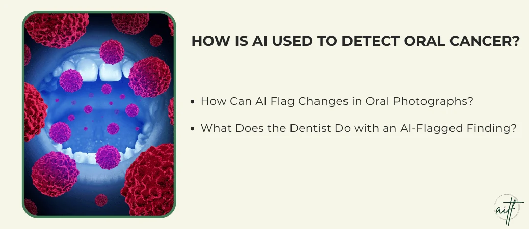
How Can AI Flag Changes in Oral Photographs?
The technology behind this is called computer vision. A program learns from thousands of labeled clinical oral photographs, some showing healthy tissue and some showing precancer or cancer.
Over time, it learns which patterns of color, texture, and shape tend to go with each group.
When your dentist takes a photo of a spot, the software compares it to what it has learned. It may draw a box around an area, give a risk score, or sort the spot into a group such as “likely harmless” or “refer for review.”
This kind of computer-aided diagnosis is meant to support early oral cancer detection, especially for small spots that are easy to overlook.
Photos also create a record. If a spot is flagged today, your dentist can compare it to a new photo in a few weeks and see whether it has grown or changed.
What Does the Dentist Do with an AI-Flagged Finding?
An AI flag is a prompt for a closer look. Your dentist treats it as clinical decision support, meaning one piece of information alongside your history, habits, and what they see and feel during the exam.
From there, your dentist may:
- Re-examine the spot and feel it for firmness or texture changes.
- Ask how long it has been there and whether it hurts or bleeds.
- Remove possible causes, like a sharp tooth edge, and recheck in about two weeks.
- Refer you to an oral surgeon or oral medicine specialist if the spot looks concerning.
A flag can also be wrong in either direction. Your dentist may decide a flagged spot is harmless, or act on a spot the software rated as low risk.
What Kinds of Data and AI Tools Are Being Studied?
Researchers are testing AI on several types of data, from simple photos to microscope slides and health records. Each one gives a different view of what is going on in the tissue.
Photographs and Other Noninvasive Imaging
Most screening research uses machine learning, and in particular deep learning. A popular type of deep learning model is the convolutional neural network (CNN), which is built to find patterns in images.
Many teams use transfer learning, where a model first trained on everyday pictures is retrained on mouth photos. This helps when medical datasets are small.
Image types being studied include:
- Clinical photographs taken with white light imaging, the same as a regular camera or smartphone.
- Autofluorescence imaging, where a special light makes unhealthy tissue look darker.
- Optical coherence tomography (OCT), which creates detailed pictures just below the surface without cutting.
- Cytology, where cells are gently brushed off a spot and examined.
Smartphone photos get a lot of attention because they are cheap and easy to share with specialists far away.
Biopsy Slides and Patient Risk Data
After a biopsy, tissue is placed on glass slides. Digital pathology scans those slides into high-resolution histopathology images, and AI can help pathologists spot abnormal cells.
Results here tend to be strong because the images show detail at the cell level. One review reported histopathology models reaching 99.26% sensitivity in a 2021 study.
Other tools use clinical data such as age, tobacco and alcohol history, and past lesions. Methods like random forests combine these factors to estimate risk.
These risk tools could help your dentist decide how closely to watch you.
How Reliable Are AI Screening Results?
AI tools have shown high diagnostic accuracy in research, but most have not been fully tested in everyday dental offices. Two numbers, sensitivity and specificity, tell you most of what you need to know about any screening test.
What Do Sensitivity and Specificity Tell Us?
Sensitivity is how often a test catches a spot that is truly a problem. Specificity is how often it correctly clears a spot that is harmless.
Here is what that looks like using one study that used intraoral photos, which found 93.9% sensitivity and 81.2% specificity:
| Measure | Result | What it means for 100 patients |
| Sensitivity | 93.9% | Of 100 true problem spots, about 94 flagged and 6 missed |
| Specificity | 81.2% | Of 100 harmless spots, about 81 cleared and 19 false alarms |
A missed lesion is the bigger worry, since it can delay care. A false alarm causes stress and extra visits, but follow-up usually sorts it out.
Systematic reviews and meta-analyses pool many studies to give a fuller picture, and they show wide ranges. OCT-based tools, for example, reported sensitivity from 75% to 100% depending on lesion type.
Why Might a Research Result Not Hold Up in a Dental Office?
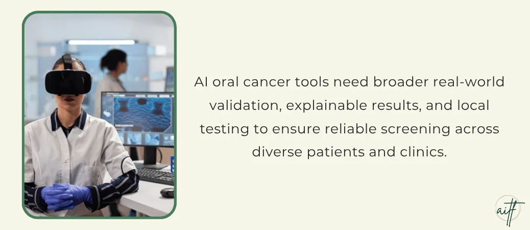
Many models are trained on photos from one hospital. Real offices use different cameras, lighting, and patient groups.
External validation tests a model on outside data, and in that same review, only four of ten included studies did it.
Researchers use data augmentation, such as flipping or brightening images, to make training sets bigger. That helps, though it cannot fully stand in for real variety.
Model interpretability also lags. Explainable AI tools show which part of a photo drove the result, so your dentist can judge whether it makes sense, but few studies use them.
Tools designed for resource-limited settings show promise for reaching more people. Each still needs testing where it will be used.
What Happens If a Dentist Finds a Suspicious Spot?
A suspicious spot leads to more checks, and a biopsy is the step that gives a real answer. Most flagged spots are not cancer, but you should never skip the follow-up.
When Is a Biopsy or Specialist Referral Needed?
Your dentist will usually recommend a biopsy or referral when:
- A sore or patch has not healed after two to three weeks.
- A patch is red, or red and white mixed.
- The spot feels hard, fixed in place, or is growing.
- You have high-risk habits and a new lesion appears.
A biopsy removes a small piece of tissue, usually with local numbing. An oral pathology specialist then reviews it under a microscope.
That histopathological diagnosis tells you whether the spot is harmless, precancerous, or oral squamous cell carcinoma, the most common type of oral cancer.
If you notice a sore that won’t heal between visits, it is one of the signs you need to see a dentist ASAP.
Why Can’t AI Confirm an Oral Cancer Diagnosis?
AI only sees what a photo or data file shows. A photo cannot reveal what the cells look like inside the tissue, and cancer is defined by those cell changes.
An oral cancer diagnosis also guides treatment planning. Your care team needs to know the exact type, how deep it goes, and whether it has spread. Those answers come from biopsy results, imaging, and specialist exams.
AI may help organize that information, but trained clinicians confirm the diagnosis and choose the treatment.
AI Can Support Screening, but Dentists Make the Decisions
AI is a promising helper for early diagnosis of oral cancer, and your dentist stays in charge of every decision. It can point to a spot, sort risk, and keep a photo record. It cannot feel a lump, hear your history, or confirm cancer.
Research shows strong numbers, yet many tools still need testing on wider groups of patients before they are routine.
For now, the best oral cancer detection comes from a careful exam, honest talk about your habits, and quick follow-up on anything unusual. AI-based clinical decision support adds to that process without replacing it.
What you can do today:
- Keep up with regular dental exams, even if your teeth feel fine.
- Tell your dentist about tobacco, alcohol, and any spot that lasts more than two weeks.
- Ask whether a photo can be taken so changes can be compared at your next visit.
- If a biopsy is recommended, schedule it promptly and ask when results will be ready.
Frequently Asked Questions
Can a dentist see signs of oral cancer during a routine exam?
Yes, many early signs are visible or can be felt during a routine exam. Your dentist checks your mouth, tongue, and neck for patches, sores, and lumps.
Some changes are subtle, which is why follow-up on any unusual spot is important.
What are the very first signs of oral cancer?
The earliest signs are often a white or red patch, or a sore that does not heal within two weeks. These spots are usually painless at first. Numbness, a lump, or unexplained bleeding can also appear.
Can AI detect oral cancer from a photo?
AI can flag a spot in a photo as suspicious, but it cannot confirm cancer. It looks for patterns linked to precancer or cancer and points them out for your dentist to review.
Accuracy varies by tool and has not been widely proven in everyday offices.
Can an AI screening result replace a biopsy?
No, an AI result cannot replace a biopsy. Only a tissue sample reviewed by a pathologist can show whether cells are cancerous. AI may help decide which spots need a biopsy sooner.
How long can oral cancer go undetected?
Oral cancer can go unnoticed for months because early spots often cause no pain. That delay is one reason so many cases are found late.
Regular exams and quick follow-up on lasting sores give you the best chance of catching it early.